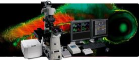
The HT-2 series opens a new era of 3D correlative imaging, combining the holotomography and fluorescence methods.
The HT-2 allows the viewing of a 3D RI tomographic image of a living cell with minimal damage, while at the same time targeting consumer interest by allowing for fluorescence labeling in the vary same cell.
Research and diagnosis in the life sciences depends on the information that can be found using cellular analysis. However, current microscopy techniques limit the quantity and quality of information available to researchers and clinicians
The HT-2, a microscope that displays both 3D holograms and 3D fluorescence imaging, is able to overcome these limitations through use of new and existing technology. Its abilities can enable researchers and clinicians to break into new frontiers of understanding, diagnosing, and treating disease.
HT-2 is the world’s first microscopy combining both holotomography and 3D fluorescence imaging in one unit.
The HT-2 is an update to our HT-1 microscope. The HT-1 microscope uses holotomographic technology to measure the 3D and 4D refractive indexes using a dynamic micromirror device (DMD) to add unprecedented stability and precision to the performance of the microscope. These innovations allow for quantitative, label free imaging of cells. The HT-2 has all the capabilities of the HT-1, but combines the holotomographic imaging capabilities of the original version with the added ability to perform fluorescence microscopy.