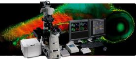
ER: Enhance Your Confocal Resolution
The A1 ER enhanced resolution Module combines powerful GPU-based processing and specialized deconvolution algorithms to dramatically enhance the spatial resolution of your A1+ or A1R+ confocal microscope with minimal processing time.
The A1 ER Module provides high quality PSF models for Nikon’s high performance objective lenses, taking the guesswork out of deconvolution analysis. Other features include automatic or manual mode for iteration selection, enhanced spherical aberration correction, and robust algorithms for noise estimation and removal.
Galvano Scanner Enables High-resolution Imaging
The A1+ utilizes a galvano scanner which enables high-resolution imaging of up to 4096 x 4096 pixels. In addition, with the newly developed scanner driving and sampling systems, plus image correction technology, high-speed acquisition of 10 fps (512 x 512 pixels) is also possible.
Zebrafish labeled with four probes (captured with galvano scanner) Nucleus (blue): Hoechst33342, Pupil (green): GFP, Nerve (yellow): Alexa555, Muscle (red): Alexa647. | Drosophilasp. embryonic heart | Bovine brain microvascular endothelial cells labeled with MitoTracker (mitochondria, yellow), phalloidin (actin, blue) and Hoechst (DNA, magenta). |
GaAsP Multi Detector Unit
Nikon developed the GaAsP multi-detector unit equipped with two GaAsP PMTs and two normal PMTs.
A GaAsP PMT has much higher sensitivity than a normal PMT, thus acquisition of brighter signals with minimal background noise is possible with a GaAsP PMT, even with weak fluorescence, which, until now, has been difficult to detect.
When using resonant scanners, the GaAsP PMT enables low-noise, high-speed imaging.

Sensitivity comparison of GaAsP PMT and normal PMT GaAsP PMT realizes higher sensitivity than normal PMT, thus offering high quantum efficiency up to 45%. * Quantum efficiency indicates logarithm
Increased Light Detection Efficiency
The low-angle incidence method utilized on the dichroic mirrors increases fluorescence efficiency by 30%.
Conventional 45° incidence angle method |

Reflection-transmission characteristics have high polarization dependence Low-angle
incidence method

Reflection-transmission characteristics have lower polarization dependence
By employing the hexagonal pinhole, higher brightness equivalent to that of a circular pinhole is achieved.
64% of the area of the circle | 30% more light |


Hexagonal pinhole
83% of the area of the circle
Nikon’s original dual integration signal processing technology (DISP) has been implemented in the image processing circuitry to improve electrical efficiency, resulting in an extremely high S/N ratio.
Enhanced Spectral Imaging
Acquisition of a 32-channel spectral image (512 x 512 pixels) with a single scan in 0.6 second is possible. Moreover, 512 x 32-pixel images can be captured at 24 fps.
Accurate, High-speed Unmixing
Accurate spectral unmixing provides maximum performance in the separation of closely overlapping fluorescence spectra and the elimination of autofluorescence. Superior algorithms and high-speed data processing enable real time unmixing during image acquisition.

Spectral and unmixed images of five-color-fluorescence-labeled HeLa cells. Specimen courtesy of: Dr Tadashi Karashima, Department of Dermatology, Kurume University School of Medicine
V-filtering Function
Filter-less intensity adjustment is possible by selecting desired spectral ranges from 32 channels that match the spectrum of the fluorescence probe in use and combining them to perform the filtering function.

Increased flexibility and ease of use
NIS-Elements C control software enables integrated control of the confocal imaging system, microscope and peripheral devices with a simple and intuitive interface. Diverse reliable analysis functions are also available.