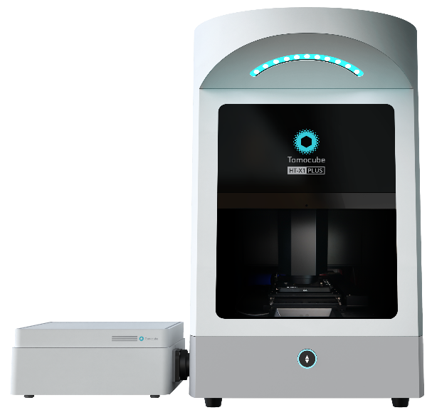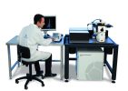

The Tomocube HT-X1 Holotomography system delivers advanced, label-free 3D and 4D quantitative live-cell imaging using a low-coherence light source. This innovative approach provides improved resolution while eliminating laser-induced speckle noise, enabling researchers to observe living cells with greater clarity and accuracy.
The HT-X1 Holotomography system is compatible with diverse multi-well plates and supports a wide range of biological samples. It can image confluent cell sheets, individual cells and thick specimens while maintaining detailed 3D visualization.
Beyond visualization, the HT-X1 provides quantitative information about cells and intracellular structures. Researchers can measure parameters such as cell volume, surface area and dry mass, helping to generate meaningful quantitative data from label-free live-cell imaging.
Traditional cellular imaging often relies on fluorescent or other labels to visualize specific molecules and structures. Although labeling techniques can provide valuable information, the labeling process may affect cells and interfere with their natural biological behavior. Fluorescence imaging can also expose cells to intense illumination, which may contribute to phototoxic effects during extended experiments.
Holotomography (HT) offers a label-free approach to live-cell imaging. Instead of relying on fluorescent labels, the technology captures the intrinsic light-scattering properties of biological samples using very low light intensity.
The HT-X1 Holotomography system measures the refractive index (RI) distribution within cells and their intracellular structures. This information can then be reconstructed into detailed 3D images and selectively pseudo-colored for clearer visualization of cellular components.
Because Holotomography captures quantitative refractive index information, the HT-X1 can also provide valuable 3D quantitative cell measurements, including volume, surface area and dry mass. Researchers can therefore examine both cellular morphology and quantitative characteristics within the same imaging workflow.
The HT-X1 enables researchers to move beyond conventional 2D observation toward 3D and 4D live-cell imaging. By recording cellular structures over time, researchers can visualize changes in morphology, growth and intracellular organization while reducing the need for fluorescent labeling.
This makes the HT-X1 suitable for applications where maintaining natural cell behavior and obtaining quantitative measurements are important.
The low-coherence light source used by the HT-X1 provides improved imaging performance while reducing laser-induced speckle noise. This helps researchers obtain clearer images of cellular structures and improves the visualization of complex biological samples.
The HT-X1 supports imaging across a variety of multi-well plates, making it suitable for research workflows that require analysis of multiple samples. Its compatibility with multi-well formats can support higher-throughput experiments while maintaining label-free quantitative imaging capabilities.
Holotomography provides more than visual images. The HT-X1 captures quantitative information that can be used to analyze individual cells and their intracellular structures.
Depending on the imaging workflow, measurable parameters can include:
These quantitative measurements can help researchers track cellular changes and compare biological samples over time.
The HT-X1 is designed to work with diverse biological specimens, including individual cells, confluent cell sheets and thick samples. This flexibility allows researchers to explore a broad range of biological structures without relying exclusively on fluorescent labeling.
With label-free 3D and 4D imaging, quantitative analysis, multi-well plate compatibility and low-speckle imaging, the Tomocube HT-X1 Holotomography system provides a powerful platform for advanced live-cell research.
Discover how HT-X1 can expand your approach to cellular imaging and quantitative biological analysis.